





- A +
- A
- A -
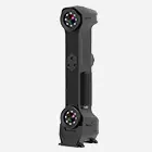
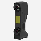
As precision components designed to interface with the human body, the dimensional accuracy of medical joints directly impacts post-implantation fit and operational safety. The client required 3D data acquisition of these joints to control production quality by detecting deviations through 3D comparisons against the manufacturing model. Given the intricate surface features of the joints and the client's stringent requirements for both scanning speed and accuracy—which traditional measurement methods could not meet—the HOLON 3D smart 49 handheld 3D scanner was selected to provide precise data support for quality control and personalized product development.

Photograph of a medical joint model

On-site scan image
Medical joint surfaces feature intricate textures, varying curvatures, and mating grooves—details that directly influence how well the component fits the human body. Traditional measurement tools struggle to capture these features precisely, often resulting in ambiguous or incomplete data that fails to meet high-precision inspection standards. Manual point-by-point measurement is extremely inefficient; the excessive time required for data acquisition on a single component neither aligns with the need for rapid quality control nor supports the timely progress of subsequent personalized design efforts. Furthermore, contact-based measurement risks abrading the surface and damaging fine features. Additionally, the manual processing required to prepare traditional measurement data for comparison and analysis introduces complexity and potential errors, severely compromising the reliability of inspection results and the accuracy of subsequent research.

Scan data plot

Scan data plot
The HOLON 3D Smart 49 handheld 3D scanner enables efficient and precise 3D data acquisition for medical joint components. With its ultra-high precision, the scanner clearly captures intricate surface textures, curvature variations, and mating grooves, accurately reproducing the component's true form to meet requirements for deviation analysis against manufacturing models while providing high-fidelity data for subsequent studies on anatomical fit. Its high-speed scanning capabilities drastically reduce data acquisition time and rapidly capture full-surface information, satisfying the client's stringent efficiency demands. The non-contact scanning method avoids physical contact with the joint surface, effectively protecting delicate features from damage. Furthermore, the flexible handheld operation allows for easy coverage of complex structures and hidden areas, ensuring comprehensive data acquisition without blind spots. The resulting 3D data is complete and precise, fully meeting the client's needs for deviation analysis and preliminary research into anatomical fit, thereby providing a reliable digital foundation for quality improvement and personalized development of medical joint components.

Reverse modeling data diagram

Reverse modeling data diagram